Salivary stone disease: causes, symptoms, consequences and treatment
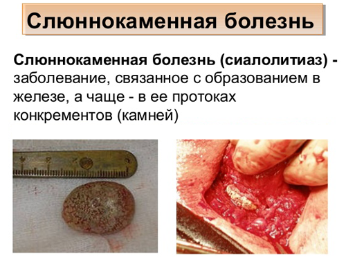
Salivary stone disease is a pathological process in which a dense mineral formation is formed in the salivary gland - usually in the duct, less often in the parenchyma - salivolith, it is also a calculus. Its composition is close to the composition of tartar, sizes can vary from several millimeters to several centimeters. A person usually does not notice their appearance until the stone grows in size so much that it covers the lumen of the salivary gland, which leads to severe pain.
The choice of treatment - medication or operable - depends on the stage of the process, the size of the stone in the duct of the salivary gland, its exact location and other circumstances.
Content
Reasons for the appearance
The exact mechanism for the formation of salivary stones has not yet been explained by science. However, doctors have identified a number of pathogenic factors that can lead to salivary stone disease.
The causes of stones in the salivary gland can be:
- avitominosis (especially vitamin A deficiency);
- disturbances in the metabolism of phosphorus and calcium;
- urolithiasis disease;
- hyperparathyroidism;
- hypervitaminosis D;
- gout;
- diabetes;
- getting into the duct of a foreign body (a solid particle of food, a fragment of a tooth, etc.);
- pathology of the ducts;
- mechanical injuries;
- consequences of wearing crowns.
A combination of several causes leads to this rather rare disease. Additional aggravating factors are bad habits, especially smoking, an insufficient level of oral hygiene, etc. The appearance of a stone in the duct of the salivary gland may be facilitated by the use of certain drugs:
- means for lowering pressure;
- diuretics;
- psychotropic;
- antihistamines.
Directly to the formation of a stone is the leaching of mineral substances from saliva, the deterioration of its properties, as well as a shift in the acid-base balance towards alkalis (which explains the constant unpleasant aftertaste in the mouth). In combination with a narrowing of the lumen of the duct, this leads to clogging with a dense mass, prone to solidification: this is how the stone forms in the salivary gland.
Stone composition and location options
Stones of the salivary glands are dense formations of a yellowish-white or yellow shape, with a tuberous surface. Composition - mineral-organic. The nucleus can be one of two types: either of a microbial nature, which is a colony of special bacteria - actinomycetes, or it can be a desquamated and cornified epithelium and / or some kind of foreign body entering the duct.
Around a foreign body - a fragment of a tooth, a fish bone that got there during a meal, a hair from a toothbrush, etc. - a layer of organic and inorganic deposits gradually grows, turning into a complex natural composite. Organics in it can be up to 30%, mainly these are particles of the epithelium, mucin and amino acids. Inorganic components may be:
- calcium salts;
- sodium;
- magnesium;
- potassium;
- iron;
- chlorine etc.
In the case of the bacterial nucleus, everything is somewhat more complicated. A large stone in the duct of the salivary gland is always accompanied by an infection and an inflammatory process, however, the question of what was the root cause - infection or stone formation - remains open.
Please note: the mass of a solid formation can vary from 3 to 30 g, and over time, the stones tend to increase.
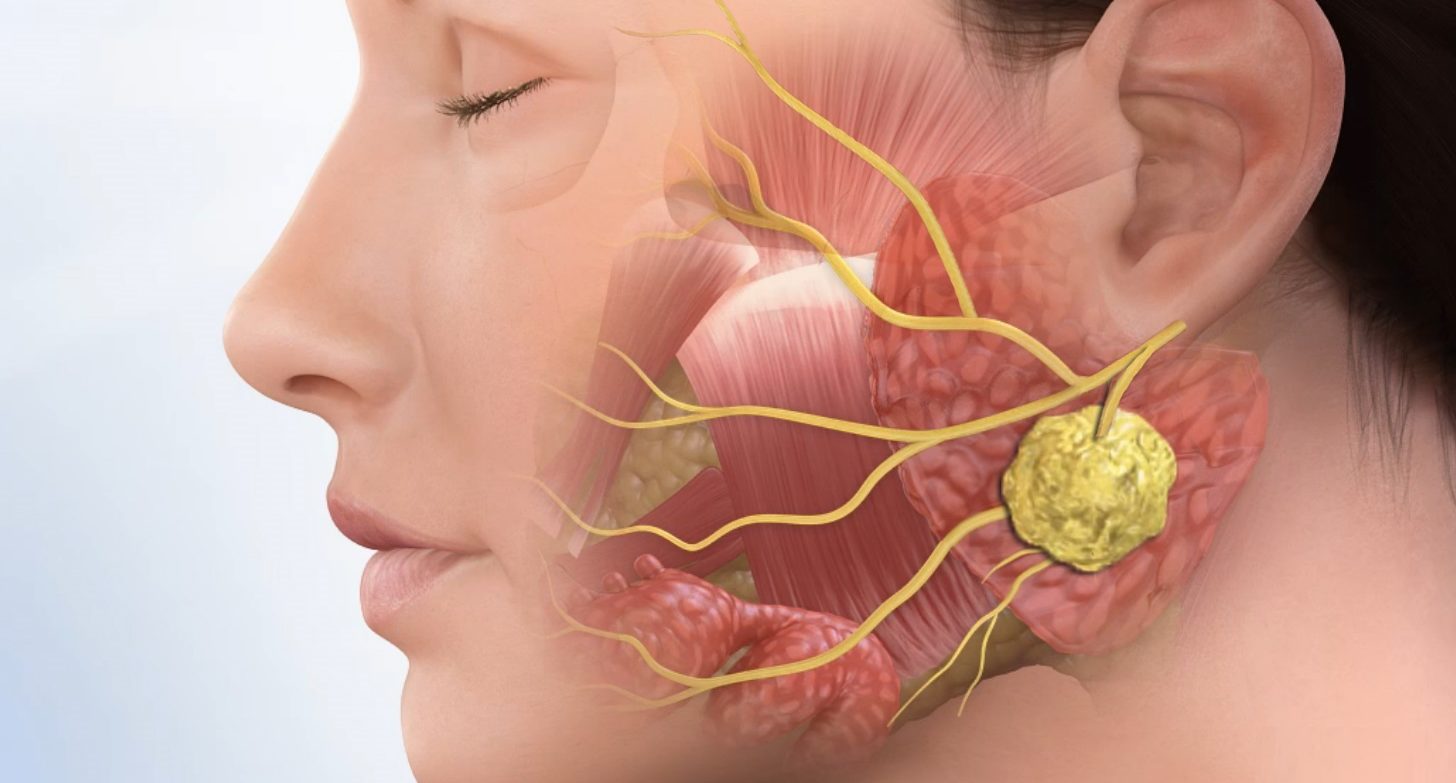
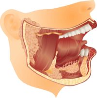
Salivary stone disease, which reached the stage of the inflammatory process, is called sialolithiasis. Salivary gland stones are most common in the submandibular gland, in about 8 out of a hundred cases in the parotid gland, and very rarely under the tongue and in the small salivary glands - labial, buccal, etc. The disease can have an acute and chronic form.
Symptomatology
The patient usually does not notice the formation of stone in the parenchyma until he completely blocks the duct. After this, pain may occur, acute, but in the form of short-term attacks - the so-called. salivary colic. The attack can last about 20 minutes.
Symptoms of stones in the salivary gland differ from the nature of the disease - whether it occurs in acute or chronic form. The acute form implies a pronounced pain syndrome, bouts of colic, as well as the following symptoms and consequences:
- a feeling of fullness in that part of the oral cavity where the stone formed;
- frequent pain when eating;
- bad taste in the mouth;
- the appearance of an abscess or phlegmon in the oral cavity;
- soreness with pressure and / or palpation;
- increase in body temperature to 37.5 degrees;
- general weakness;
- headaches;
- opening the entrance to the salivary canal with the release of pus from the opening;
- very small amount of saliva, dry mucous membranes.
With sialolithiasis, it is painful to move the jaw while eating. It is painful to swallow, and the pain is given to the region of the ear or temple, as well as to the throat and tongue (with sialolithiasis of the submandibular gland).
Please note: an acute form of the disease can develop within a few hours. Especially severe pain occurs if the stone independently leaves the gland in soft tissue.
In a chronic form, a stone in the duct of the salivary gland may not show itself except for the following symptoms: swelling of the neck and face, constant tension of the facial muscles, as well as swelling in the area of the affected gland due to the increase in size. Pain as a symptom in a chronic form may be mild or not appear at all.
Diagnostics
Salivary stone disease should be differentiated from other diseases of the oral cavity that cause similar symptoms (fever, pain when swallowing, swelling). It can be:
- various tumors of the oral cavity;
- periandibular phlegmon;
- lymphadenitis;
- abscess.
Diagnosis of a stone in the salivary gland duct is carried out by a dentist, in the absence of it, by a general practitioner. The first step is visual inspection and palpation - in some cases, the stone can be seen or, when palpated, determine its location. The gaping salivary canal and the pus that stands out from it lends itself to visual detection.
If it is not possible to visually detect the stone, then the diagnostic methods will depend on the form and stage of the disease. Most commonly assigned studies:
- radiography;
- sialography;
- sialoscopy;
- Ultrasound of the salivary glands;
- biochemical analysis;
- CT scan.
The choice of a specific set of research and diagnostic measures is at the discretion of the doctor. Much depends on the location of salivolitis, the speed with which you need to obtain data, the necessary accuracy and the diagnosis of possible concomitant diseases.
 So, if the disease is in a chronic form and pain is absent, then the doctor can use a special probe to study the salivary canal, to determine the size of the mouth and the depth of salivolitis. If the stage is acute, then most often a set of diagnostic tools is used from X-ray and sialography (contrast x-ray), as well as ultrasound. In more complex cases, if an X-ray is of little use, a computer tomograph comes into play.
To establish the nature of the inflammatory process, a saliva cytogram is used, as well as a general biochemical analysis.
The average cost of an ultrasound of the salivary glands in Moscow is 1200 r, an x-ray of the oral cavity is 1250 r.
Methods of treatment for salivary stone disease
Treatment options for salivary stone disease will depend on what the diagnostic methods show. In some cases, removing a stone from the salivary gland by a doctor may not be necessary: small, up to 2-3 mm, stones can be washed out of the ducts with saliva.
Treatment of sialolithiasis can be either conservative - medication, or surgical. The general case may involve the combination of two methods, especially if there are several calculi (approximately 25% of all situations). With the help of surgical intervention, a large stone is removed from the salivary gland, and small medications are obtained by medical methods. This method can be applied so as not to expand the wound beyond what is necessary.
Drug treatment is used to anesthetize and stop the inflammatory process.
Conservative methods
Treatment of a stone in the salivary gland with drugs has two directions: the first helps to reduce pain and treat the inflammatory process, and the second involves the administration of salivary preparations, which cause excessive salivation and leaching of small stones. Thus, the following drugs are used:
 non-steroidal anti-inflammatory drugs to reduce pain, eliminate inflammation and reduce swelling;
- antibiotics to fight bacterial infection;
- salivary preparations.

Among the latter can be noted Kanefron, potassium iodide, pilocarpine hydrochloride.
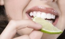
Also, the patient is additionally prescribed a diet that enhances the secretion of the salivary glands. It consists of acidic products, which in themselves have increased salivary properties and normalize the acid-base balance, which, with sialolithiasis, is “knocked down” to the alkaline side. Some acids (for example, citric acid) have the ability to destroy salivolites.
The patient must include in the diet a large number of beets, sauerkraut, squash and cranberries. You can drink a decoction of rose hips or knotweed herbs, dissolve a slice of lemon, rinse your mouth with a solution of salt and soda.
To stimulate the destruction and excretion of calculus with saliva, the doctor can use the effect on the affected gland with weak electric current discharges that do not affect the whole body.
Surgical methods
The simplest surgical method is to remove calculus with tweezers if they are at the mouth of the canal. Lithotripsy is also used - this is the crushing of a stone using ultrasound.
If it comes to inflammation and abscess, then an operation is prescribed under local anesthesia, during which the abscess is opened and cleaned, drainage is established and the stone is removed. The wound is not sutured.
If a serious pathology of the salivary gland is detected, it is removed - extirpation.