Oral cancer: causes, symptoms, treatment and prognosis for cancer of the cheeks, palate, tongue, gums, and the bottom of the oral cavity
The human oral cavity is lined with a mucous membrane formed by epithelial cells that can transform into malignant cells - this is how cancer of the oral mucosa develops. In the general structure of cancer, this pathology ranges from 2% (in Europe and Russia) to 40-50% (in Asian countries and India). Mostly male patients over 60 suffer from it, it is extremely rare in children.
Content
Causes
The exact cause leading to the appearance of neoplasms in the mouth has not been established. The researchers only revealed a number of factors that significantly increase the likelihood of developing this disease. The key among them are bad habits - smoking, chewing nasvay or betel, as well as alcohol abuse.
Additional factors are:
- Chronic mechanical injuries of the oral cavity.
- Use of poor-quality or poorly fitted dentures.
- Poor fillings and tooth injuries - sharp edges of fillings and broken teeth cause permanent injury to the mucous membranes of the cheeks and tongue.
- Gum injury with dental tools.
- Poor hygiene.
- The use of metal prostheses from different metals in dental prosthetics - galvanic stress can occur between different metals, which leads to damage to cells and their malignancy.
According to recent studies in virology and medicine, a specific role in the development of oncology of the oral cavity belongs to human papilloma viruses, which can be transmitted by kisses.
An increased frequency of development of this pathology was noted in persons working in difficult and harmful conditions: in constant contact with harmful substances, in conditions with high or excessively low temperature and high humidity.
Exposure to hot and spicy foods also promotes the formation of tumors on the oral mucosa. The situation is aggravated by a deficiency in vitamin A and the presence of inflammation or precancerous disease in the oral cavity.
Precancerous diseases that can degenerate into cancer of the oral mucosa
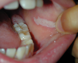
- Leukoplakia. It looks like a whitish spot on the mucous membrane in any area of the oral cavity: in the sky, on the cheeks near the lips on the inside. It is characterized by keratinization of the epithelium.
- Erythroplakia. It is characterized by the appearance of red foci, abundantly penetrated by blood vessels. Up to half of cases of erythroplakia are transformed into oncology.
- Dysplasia - actually a precancer. A study of dysplastic lesions under a microscope shows that some of the cells have already acquired the features of malignancy. If this pathology is ignored, in 99% of cases, oral cancer develops in a few months.
Symptoms and stages of oral cancer
At the very initial stage, cancer of the oral mucosa may not bother, only a part of patients feel some unusual discomfort in the mouth. On examination, you can see a crack on the mucosa, a small tubercle or a seal. About a third of cancer patients complain of unexpressed pain that masquerades as symptoms of inflammatory diseases: glossitis, gingivitis.
The progress of the disease is usually accompanied by an increase in pain, even if the inflammation has already passed. Pain can radiate to the forehead, temple, jaw. Very often, patients associate these pains with toothaches.
Untimely diagnosis allows the disease to go to an advanced stage when the following symptoms of oral cancer develop:
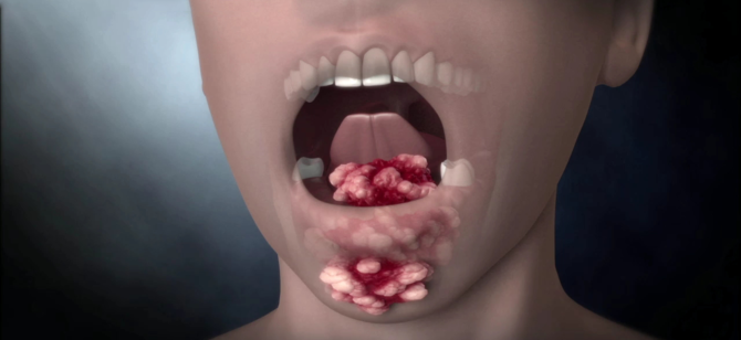
- An ulcer or growth on the mucosa appears.
- The decay of the tumor is accompanied by an unpleasant putrefactive odor.
- The pain becomes constant.
In advanced cases, the deformation of the face due to the germination of pathological tissue into the surrounding structures: muscles and bones joins the symptoms of a cancer of the mucous membrane of the oral cavity. Symptoms of intoxication are increasing: patients complain of general weakness, rapid fatigability, and nausea.
The absence of treatment with an advanced stage of cancer leads to the fact that the patient develops metastases. First, regional lymph nodes (cervical, submandibular) are affected. Then parenchymal organs can be affected - the liver and lungs. Often there is a metastatic lesion of the bones.
Classification
According to its microscopic structure, cancer of the oral mucosa is a squamous type. There are several of its forms:
- Keratinizing squamous cell carcinoma. It looks like a cluster of keratinized epithelium ("cancerous pearls"). It makes up to 95% of cases of the development of the pathology of this localization.
- Non-keratinizing squamous. It is manifested by the growth of cancerous epithelial cells without keratinization sites.
- Low grade (carcinoma). This is the most malignant and difficult to diagnose form.
- Cancer of the oral mucosa in situ. The rarest form.
Depending on the characteristics of tumor growth, the following forms are distinguished:
- Peptic ulcer is one or several ulcers that gradually grow and tend to grow and merge. Usually the bottom of the ulcers is covered with an unpleasant plaque.
- Knotty - characterized by the appearance on the mucous membrane of a dense growth in the form of a node covered with whitish spots.
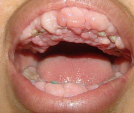
- Papillary - manifests itself as fast-growing, dense growths resembling warts. Outgrowths are usually accompanied by edema of the underlying tissues.
Individual forms of cancer of the oral mucosa
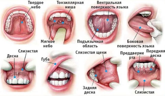
- Cancer of the tongue. A typical location of the pathology is the lateral surface of the tongue, less often the tumor is found on the root of the tongue, back or lower surface. A malignant tumor in the early stages leads to a disorder of chewing and swallowing, which facilitates the diagnosis.
-
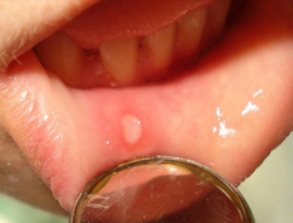
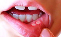
Cancer of the cheek mucosa. This tumor is often masked by aphthous ulcers located on the line of the mouth on the cheeks. An increase in the diameter of the ulcer and its germination in the masticatory muscles leads to a restriction in the opening of the mouth, which is a typical symptom of cancer of the cheek.
- Cancer of the palate and gums of the upper jaw. A rapidly growing node with a tendency to ulceration forms in the sky. An early onset of pain may be noted, especially in situations where deeper tissues are affected.
- Cancer of the mucous membrane of the bottom of the oral cavity. The first signs of the disease almost always go unnoticed. The tumor grows early in the surrounding tissue, including bone. The oncological process is often accompanied by increased salivation (salivation), dangerous development of bleeding. Tumor growth with damage to the bone tissue of the lower jaw is accompanied by facial deformation.
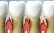
- Cancer of the mucosa on the alveolar processes. At an early stage, it is accompanied by problems with teeth - patients are worried about toothache, the teeth become loose and fall out, the gums swell. A typical phenomenon is bleeding from the socket of a fallen tooth.
Diagnostics
The diagnosis is made on the basis of patient complaints and after examination of the oral mucosa. A tumor biopsy helps confirm the diagnosis.Technological diagnostic methods, such as ultrasound or tomography, with these tumors are not very informative. To detect damage to the bone tissue of the lower and upper jaws, the patient is prescribed an x-ray of the facial skeleton.
To identify metastatic lesions, doctors usually prescribe an abdominal ultrasound and chest x-ray. Perhaps the appointment of computed or magnetic resonance imaging.
More often the first tumors in the oral cavity are noticed by dentists in connection with the characteristics of their profession. If the first signs of oncology in the mouth are detected, the patient is surely referred for an oncologist consultation.
Treatment methods
When treating tumors of the oral mucosa, doctors use the entire arsenal of available drugs:
- Radiotherapy (radiation therapy).
- Chemotherapy
- Surgery
Depending on the stage of the cancer process, both monomethodics and combined treatment of cancer are used. At stages 1 and 2 of the disease, radiotherapy gives a good effect. The advantage of this method is that after it the appearance of cosmetic or functional defects is almost completely eliminated. In addition, it is relatively easily perceived by patients and has a minimum of side effects. However, at stages 3 and 4 of the disease, the effectiveness of this treatment method is very low.
Surgical operations are in demand at stages 3 and 4 of oral cancer. The scope of the operation depends on the prevalence of the process. It is important to excise the tumor completely (within healthy tissue) to eliminate the risk of relapse. In radical surgery, it is often required to excise the muscles or resect the bone, which leads to severe cosmetic defects.
After surgery to treat tumors of the oral cavity in some cases, plastic surgery is required. For breathing difficulties, a tracheostomy (hole in the throat) may be placed on the patient.
Among all treatment methods, chemotherapy for oral cancer is the least effective, but it can reduce the volume of the neoplasm by more than 50%, which greatly facilitates the surgical operation. Since chemotherapy does not cure this type of cancer, it is used only as one of the stages of complex treatment.
In cases where a patient with an advanced degree of oncology has very little left to live due to metastases or cancer intoxication, palliative therapy comes first in the treatment. This treatment is aimed at combating concomitant complications (bleeding, pain) and consists in providing a hopeless patient with a normal quality of life. In palliative therapy, painkillers are used.
The use in the treatment of fairly aggressive methods (radiation and chemotherapy) affects the health of the patient. During the course of treatment, the following side effects from medications may be noted:
- Stool disorder in the form of profuse diarrhea.
- Constant nausea, accompanied by vomiting.
- Baldness.
- Development of immunodeficiency (patients during chemoradiation treatment should avoid acute respiratory viral infections).
During the treatment of oncopathology of the oral mucosa, patients need to fully eat - the diet should be rich in proteins of both animal and plant origin. If oral nutrition is not possible (by mouth), food can be administered via a pre-installed probe or intravenously (use special mixtures for parenteral nutrition).
Prevention
The main preventive value in the fight against cancer of the oral mucosa is the rejection of bad habits. Be sure to quit smoking, chewing betel nut, use nasvay. It is recommended to give up alcohol.
Reducing trauma to the cheeks, tongue, gums also reduces the risk of tumors of the described localization. All teeth should be cured, established fillings should be processed.If necessary, prosthetics should be very carefully selected prosthesis, so that it is convenient in operation and does not cause discomfort.
Products with irritating effects should be excluded from the diet; very hot foods should not be consumed. When the first signs and symptoms of oncology of the oral cavity appear, you should immediately consult a specialist.
To reduce the likelihood of oncology, people working in hazardous industries should actively use personal protective equipment - work clothing, respirators.
With regularity at least once a year, and when precancerous conditions are detected every quarter, you need to undergo preventive examinations at the dentist and oncologist.
Forecast
When treating cancer at an early stage, with an insignificant degree of damage to the surrounding tissues, the prognosis is very favorable - after recovery, you can live without any particular health concerns. In 80% of people with a tongue tumor who have undergone isolated radiotherapy, relapse is not recorded for 5 years. Tumors of the bottom of the oral cavity and cheeks are more unfavorable in this regard - for them, a five-year relapse-free period is observed in 60 and 70% of cases, respectively.
The larger the size of the tumor, and the more surrounding tissue it affects, the sadder the prognosis. Some patients with the fourth stage have only a few months left to live, especially if distant metastases have appeared. With surgical treatment, the prognosis may depend on the fact that there are no malignant cells left in the body after the operation, the repeated growth of which will give a relapse.

