Causes, signs and treatment of a wedge-shaped tooth defect
A wedge-shaped tooth defect can develop even in people who pay attention to hygiene and oral health. Diagnosis of the disease is complicated by the similarity of symptoms with a number of other, more common diseases. The treatment of a wedge-shaped tooth defect is complicated by the difficulty of establishing the reason why it developed. The exact etiology of the disease has not been established.
Content
Pathology Description
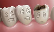
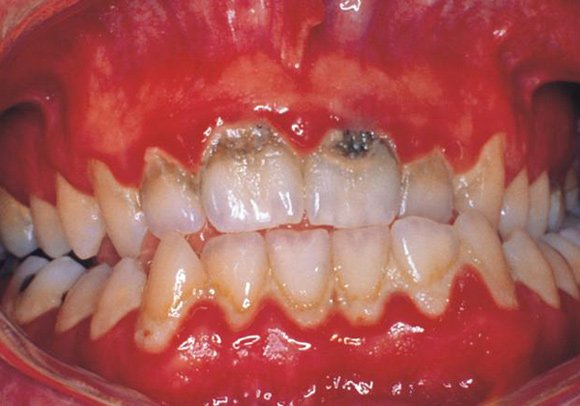
A wedge-shaped tooth defect is a lesion of the cervical region of tooth enamel, resembling a protrusion in the shape of a triangle or wedge. The characteristic form of the affected area is one of the main signs of the disease.
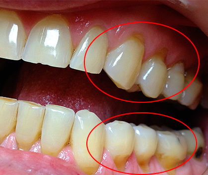
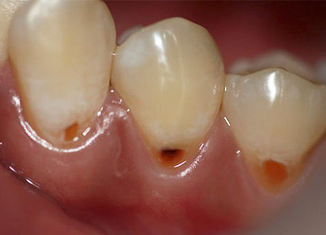
Photo: wedge-shaped tooth defect
Usually, pathology occurs on teeth that experience maximum load when chewing - fangs and premolars. In most cases, the disease affects the tissues of not one tooth, but several nearby dental units. The likelihood of developing pathology increases with age.
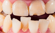
The clinical manifestations of the disease are similar to signs of cervical caries, enamel erosion and acid necrosis. However, the etiology of this disease is different, and it is treated differently than caries or other dental diseases.
Causes of a wedge-shaped tooth defect
The exact causes of the wedge-shaped defect are unknown to modern medicine. Doctors identified five main theories that explain why pathology can develop:
- Visceral. Diseases of the nervous, digestive and endocrine systems can have a negative effect on dental health. Such pathologies can lead to a violation of the acid-base balance in the body, including in the oral cavity. An increased level of acidity is fraught with a decrease in the enamel layer, which can provoke the formation of pathology.
- Erosive (chemical). The presence of acid in food and drink leads to thinning of enamel. In addition, excessive consumption of salty and acidic foods, carbonated drinks and the use of whitening hygienic pastes contribute to an increase in the fragility of enamel.
- Mechanical Incorrect hygiene and the wrong choice of hygiene items and oral care products. Too hard bristles of the toothbrush irritate and injure the gums during toothbrushing, and too soft can not completely clean the teeth of food debris. Enamel thinning is facilitated by improper plaque cleansing of dental tissues and toothbrushing immediately after consuming high acidity products.
- Load theory. Chewing load is unevenly distributed, especially if the situation is complicated by an incorrect bite.
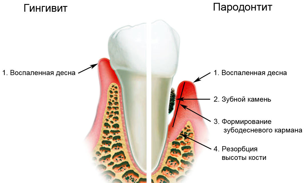
- Periodontic. Inflammation of the near-tooth tissues (gingivitis, periodontitis) can provoke the prolapse of the gingival margin. As a result, the neck of the tooth is exposed, and a wedge-shaped defect occurs.
In addition to the above, the development of pathology is facilitated by:
- Hormonal diseases, especially those that cause a violation of calcium metabolism (diabetes mellitus, osteoporosis). In women, pregnancy and lactation may be additional risk factors, accompanied by hormonal changes and leaching of calcium from the body.Also, hormonal failures can lead to stress, depression.
- Mechanical damage to teeth as a result of constant consumption of solid food, improper selection of toothpastes and brushes.
- Inadequate nutrition, leading to insufficient intake of fluoride and calcium.
- Smoking and drinking. Nicotine increases fragility of blood vessels, which entails malnutrition of periodontal tissues and their degeneration. Alcohol in large quantities leaches calcium from the body, which leads to thinning of enamel.
- Age-related changes in the body.
- Incorrect selection and inaccurate removal of braces.
- Radiation and chemotherapy.
Whether there is a genetic predisposition to the development of a wedge-shaped defect is reliably unknown. Therefore, if such a pathology was detected in one of the relatives, you should consult a doctor to determine the degree of risk, and pay attention to the prevention of the disease.
Symptoms and stages of development of a wedge-shaped tooth defect
The defect develops gradually. Each stage of the disease is characterized by special symptoms and is treated with various methods.
Defect development stages:
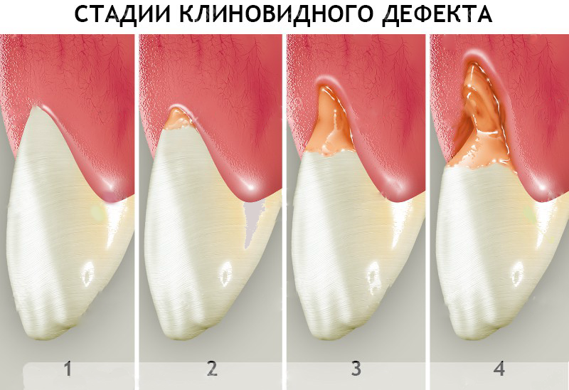
- First stage. A faint crack appears on the neck of the tooth, there is no discomfort and pain. A slight sensitivity to irritants and a slight decrease in enamel gloss may appear. It is impossible to detect a problem at home, only an attentive specialist can detect a defect during a routine dental examination.
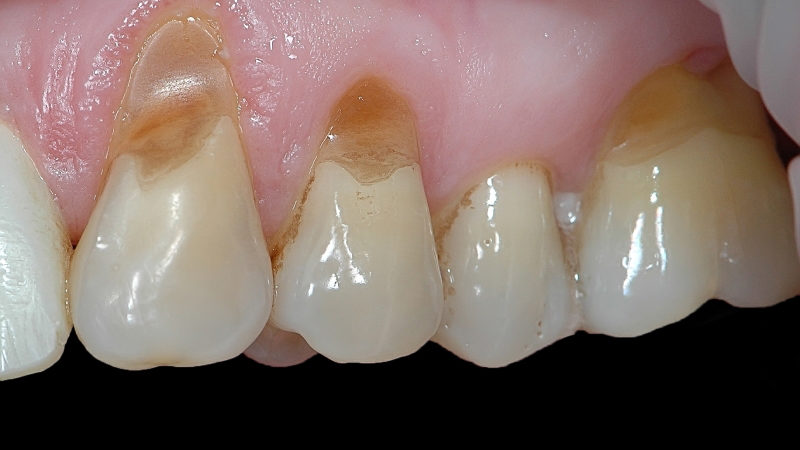
- Surface stage. A slight indentation with an area of about a millimeter appears in the surface layer of the tooth. A characteristic visual sign of the disease is the gradually increasing enamel pigmentation and the gradual exposure of the tooth neck, down to the root.
- Middle stage. The affected area increases, its depth can exceed 3 mm. Progresses the process of destruction of the tooth surface. The wedge-shaped form of the affected area with a pronounced tip is clearly visible. There is discomfort during eating, pain occurs in response to irritants and brushing your teeth.
- Deep stage. The depth of the affected area increases to 5 mm, enamel pigmentation is expressed, the dentin layer of the dental tissue is affected. The pathological process can reach the pulp, in which case sudden pain attacks appear due to inflammation of the neurovascular bundle. If the body has activated the compensatory mechanism, which led to the formation of dentin, the symptoms of the disease may be absent.
Diagnostic Methods
To cure the disease, it is important not only to detect the defect in time, but also to differentiate it from other diseases with similar symptoms, which are much more common in dental practice. As diagnostic measures apply:
- Visual inspection In the later stages, the wedge-shaped tooth defect is more easily recognized, since the wedge-shaped notch at the neck of the affected tooth reaches a solid size and is visible to the naked eye.
- Mechanical diagnosis - the effect on the affected area with irritants. The method is not always indicative, because pathology can be asymptomatic.
The difference between a wedge-shaped defect and similar pathologies
| Kind of pathology | Hard tissue erosion | Enamel Necrosis | Cervical (sphenoid) caries | Wedge-shaped tooth defect |
|---|---|---|---|---|
| Localization of the lesion | Can develop on any surface | Can develop on any surface | Develops in the cervical region of the teeth | Develops in the cervical region of premolars and canines |
| Defect bottom characteristic | Dense | The bottom is loose, the enamel layer, dentin, part of the pulp are destroyed | The fabric is soft, the edges are uneven | Enamel is destroyed, clear edges near the lesion |
| The form | Saucer-shaped | Rounded black or dark spots | Any | From the third stage, the wedge is clearly visible |
| Irritant response | Expressed reaction to cold and sweet / sour | Tooth reacts to any irritants | The affected area responds to any stimuli, painful on palpation | There may be no reaction to stimuli |
Photo of a wedge-shaped tooth defect and similar pathologies
Hard dental tissue erosion
Enamel necrosis
Cervical (sphenoid) caries
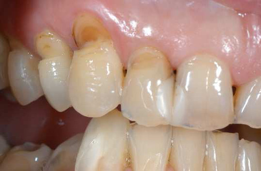
Wedge defect
Treatment of a wedge-shaped tooth defect
The goal of treatment is to repair the defect. Before treating a wedge-shaped tooth defect, the doctor must determine the degree of their defeat and reliably establish a diagnosis, excluding diseases with similar symptoms. The choice of an adequate therapeutic technique is complicated by the fact that modern science has not established the exact causes of the defect. The doctor selects treatment based on assumptions about the cause of the development of the pathology.
Possible treatment options:
- Remineralization. The method is effective at the initial stage of development of the defect. To protect against negative external influences, enamel is strengthened by means of applications with sodium gluconate solution. For treatment at home, the dentist prescribes a therapeutic paste with a high content of fluoride and calcium. Can be used at home and special gels, varnishes. The appropriate drug and duration of the course is determined by the doctor.
- Fluoridation. It is used in the later stages of the development of the disease. The doctor resorts to this type of treatment if he suspects that the cause of the pathology is problems with tooth enamel.
- Filling. It is used for a large affected area. Applied technologies and filling materials differ from those used when installing seals to eliminate carious cavities. Filling is complicated by the small area and inaccessibility of the affected area. The procedure is painful and is not a solution to the problem in the long run - the structures do not last long, because it is almost impossible to secure the seal without resorting to drilling. During chewing food, a constant increased pressure will be applied to the filling, squeezing it out of the cavity.
- Installation of veneers. Veneers are called ceramic pads that cover the external tooth surface. Their installation is recommended for the treatment of a wedge-shaped defect of 3-4 degrees. Veneers are installed after filling and additional fluorination of enamel. Pads prevent the defect from progressing and look very aesthetically pleasing.
- Prosthetics. With deep tooth decay, an artificial all-ceramic or ceramic-metal crown can be installed to return the dental unit to its original shape. Additionally, a fluorination procedure is carried out.
- Laser therapy to strengthen enamel. Painless procedures help strengthen the enamel, reduce its sensitivity and prevent further destruction of dental tissues. This method of therapy is suitable for allergic patients, pregnant women, nursing women.
If the cause of the development of the defect is an incorrect bite, it is necessary to correct it with the help of plates or braces. Without correcting the bite, it is pointless to treat the defect - the disease will return.
Disease prevention
Given the complex etiology of the development of the defect, all actions aimed at maintaining the health of teeth and gums can act as preventive measures. But the main task of the patient is to undergo scheduled examinations in dentistry at least once every six months and if any unpleasant symptoms occur.