Computed tomography of teeth in dentistry: basic information about the procedure
CT of teeth is one of the dental studies used to determine infectious and non-infectious dental pathologies. It is used in implantology, surgery, orthodontics and therapy.
Content
- Advantages of computed tomography of teeth
- Indications for dentography
- Contraindications to CT of the dentition
- The harm of computed tomography for the body
- Preparation of the oral cavity and teeth for computed tomography
- Computed tomography in dentistry
- OMS computed tomography of the oral cavity
- What is an oral tomogram?
Advantages of computed tomography of teeth
The use of computed tomography in the study of the teeth of any person has many advantages, compared with radiography and conventional visual inspection of the oral cavity:
- high speed of receiving answers to all questions of a doctor;
- small error in the study;
- high quality images, including a three-dimensional model of the jaw;
- quick determination of the source of any pain.
Thanks to computed tomography of teeth, dentists can:
- draw up a clear plan for implantation;
- choose the most suitable material for the manufacture of implants;
- control all stages of dental treatment;
- detect a neoplasm in the jaw at an early stage when the tumor can still be cured.
CT reduces the cost of treatment and prosthetics due to the maximum accuracy of the study. Thanks to computed tomography, the doctor can use the most appropriate treatment protocol, because he sees the full clinical picture of the disease.
Indications for dentography
Computed tomography of the jaw is indicated for the following pathologies:
- trauma to the jaw, teeth, joints, since it is with CT that it is possible to identify hidden injuries;
- various anomalies in the structure of the maxillofacial bones, including malocclusion;
- suspected presence of chronic inflammatory foci;
- complex tooth extraction;
- the need for complex prosthetics;
- the presence of pathologies of the dental canals.
CT of the oral cavity can be prescribed in the presence of any incomprehensible or difficult to identify symptoms.
Contraindications to CT of the dentition
Computed tomography of teeth cannot be performed at all trimesters of pregnancy. During lactation, CT is done only for strict medical reasons. Moreover, a woman should stop breastfeeding for a day or two.
This type of research is not recommended for young children and those who have recently undergone severe surgery. Dental clove diseases are safer to diagnose with x-rays.
CT of the teeth using a contrast medium is contraindicated in the following pathologies:
- hepatic and renal failure;
- pathology of the pancreas;
- diabetes;
- allergy to iodine and other components of contrast agents.
The harm of computed tomography for the body
Compared with radiography, with a tomography of the tooth jaw, a small exposure of a person occurs. During radiography, the patient receives a radiation dose equal to 0.02 mSv, with MRI even more, and with CT - 10 times less. A person is irradiated much more when he performs actions not related to medicine and dentistry:
- makes a flight in an airplane (0.09 mSv);
- passes a digital detector at the airport (0.05 mSv);
- just lives (the average annual radiation dose is 2.2 mSv).
Therefore, it is impossible to undermine health in one CT scan.A much greater danger entails the rejection of this procedure. A tumor in the early stages is in most cases curable, but delayed therapy can lead to numerous negative consequences and even death.
If a doctor prescribes a patient to undergo a CT scan, an MRI (magnetic resonance imaging) or an OPTG (orthopantomogram) to examine the teeth, do not refuse. The radiation dose received during these procedures is minimal and safe for the average adult.
Preparation of the oral cavity and teeth for computed tomography
MRI and computed tomography of teeth do not imply any limitations or special training. The patient can eat and drink even on the day of the session. But because of the features of the procedure, people with mental disorders, neurosis or claustrophobia need to take appropriate tranquilizers before CT.
Sometimes computed tomography is performed using contrast. In this case, any patient will need some preparation:
- laboratory diagnosis of KLA in order to detect an allergy to a contrast medium;
- examination of the urinary system to determine any pathologies of the kidneys.
Computed tomography in dentistry
Before research, a person needs to remove metal objects, jewelry, belts and other accessories. If the patient has braces, then the dentist removes them. Such measures are necessary to improve the accuracy of the study.
The procedure can be performed while standing, sitting or lying down. The choice of position depends on the type of pathology, the age of the patient and his physiological characteristics. In the presence of claustrophobia or CNS diseases, it is better to conduct a standing examination.
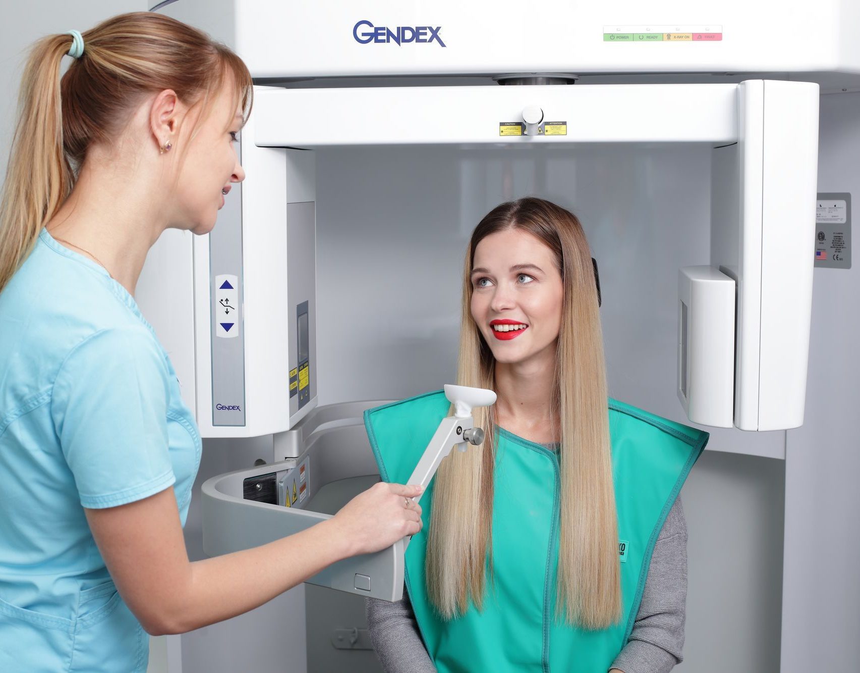
Tomograph
If necessary, a contrast agent is administered to a person. When carrying out the procedure in a supine position, a special protective apron is put on the patient. If CT is done differently, then the head of a person must be fixed with the help of a hardware rack.
Then the doctor starts the tomograph. In the course of work, he makes a faint humming noise, but you should not get up as soon as he subsides - you need to wait for the permission of the doctor. The procedure takes only a few minutes.
Often, after computed tomography using contrast, pressure rises, nausea occurs. You should notify your doctor about any symptom that manifests itself, but usually such adverse reactions are a natural response of the body to the administration of a potent drug.
OMS computed tomography of the oral cavity
In dentistry, CT can be done for free under the compulsory medical insurance policy.but only if there is appropriate evidence. To do this, you need:
- Attach to the dental clinic by compulsory medical insurance;
- get a quota in the clinic (paperwork takes 2-3 weeks);
- apply for a referral to a medical center or hospital assigned to the clinic;
- line up for examination.
To undergo a CT scan, you will need to provide:
- compulsory medical insurance policy;
- passport of a citizen of the Russian Federation or any other identification card (to be specified in the registry);
- compulsory pension insurance policy (if any);
- direction to research.
The queue in the hospital can be scheduled for a month. An examination without a queue is possible only for serious reasons, when urgent diagnosis is required. Such indications include:
- tumor neoplasms;
- head injuries;
- strokes and hemorrhages;
- spinal injuries and other serious pathologies of a similar plan;
- tuberculosis;
- sharp pain.
What is an oral tomogram?
A CT scan of the teeth is a 3D image of the jaw, obtained using hardware positioning and the use of a new fixation system at three points. In the picture, the dentition is viewed in different projections. The left side of the face in the picture is on the left and is indicated by the letter L, and the right side is on the right and is indicated by the letter R.
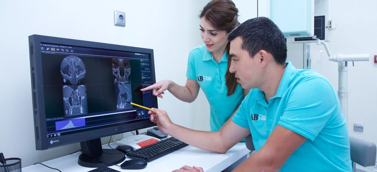
A computer tomogram of the teeth allows you to see absolutely all the defects and structural features of the jaw bones. An approximate result of computed tomography is shown in the photo:
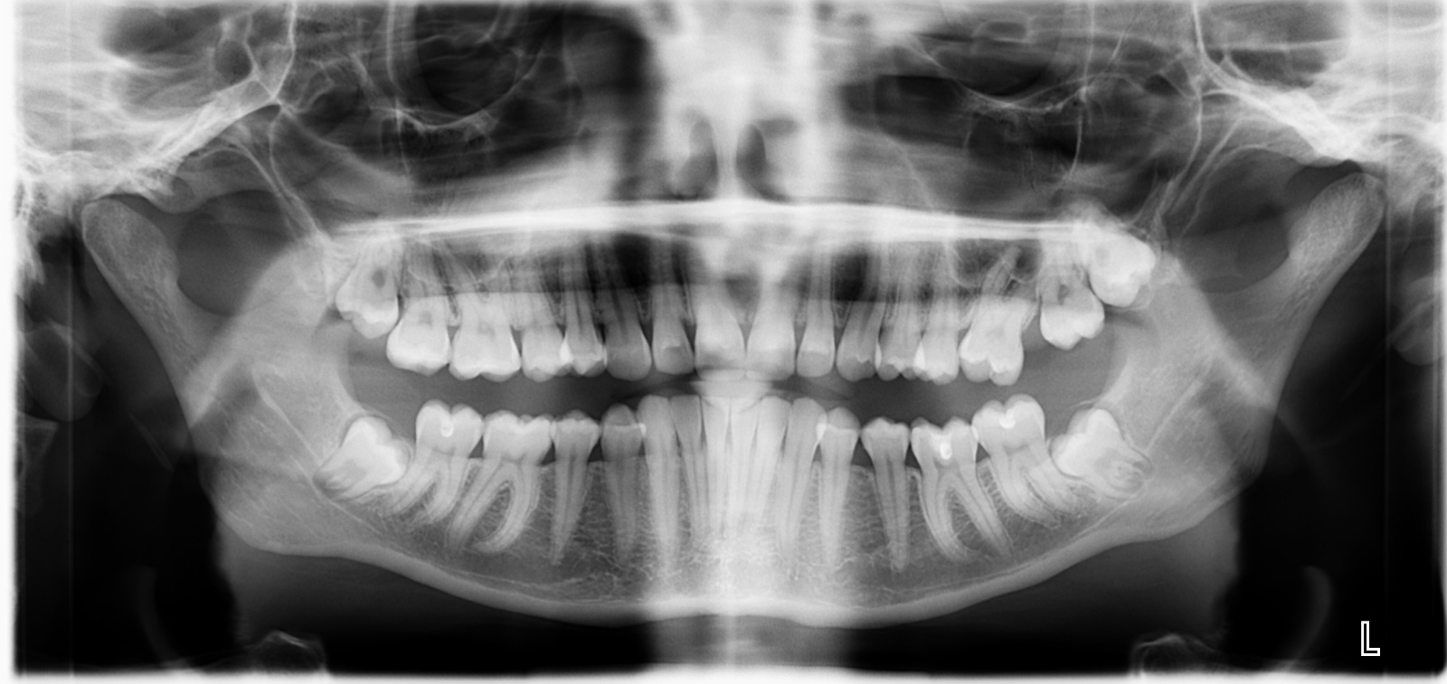
A high-quality tomogram allows even a layman to see:
- the location of the roots of the teeth, canals and primordia of uncut molars (most often the third molar);
- fillings and crowns;
- defects in soft and hard tissues of the oral cavity and jaw;
- condition of the nasal passages;
- maxillary sinuses;
- temporomandibular joints.
Computed tomography allows you to identify various pathologies of the jaw and teeth. CT of the dentofacial system can even be done according to the compulsory medical insurance policy, but in this case you will have to wait about a month to get a picture.