Panoramic picture of teeth or orthopantomogram: what is it, what shows
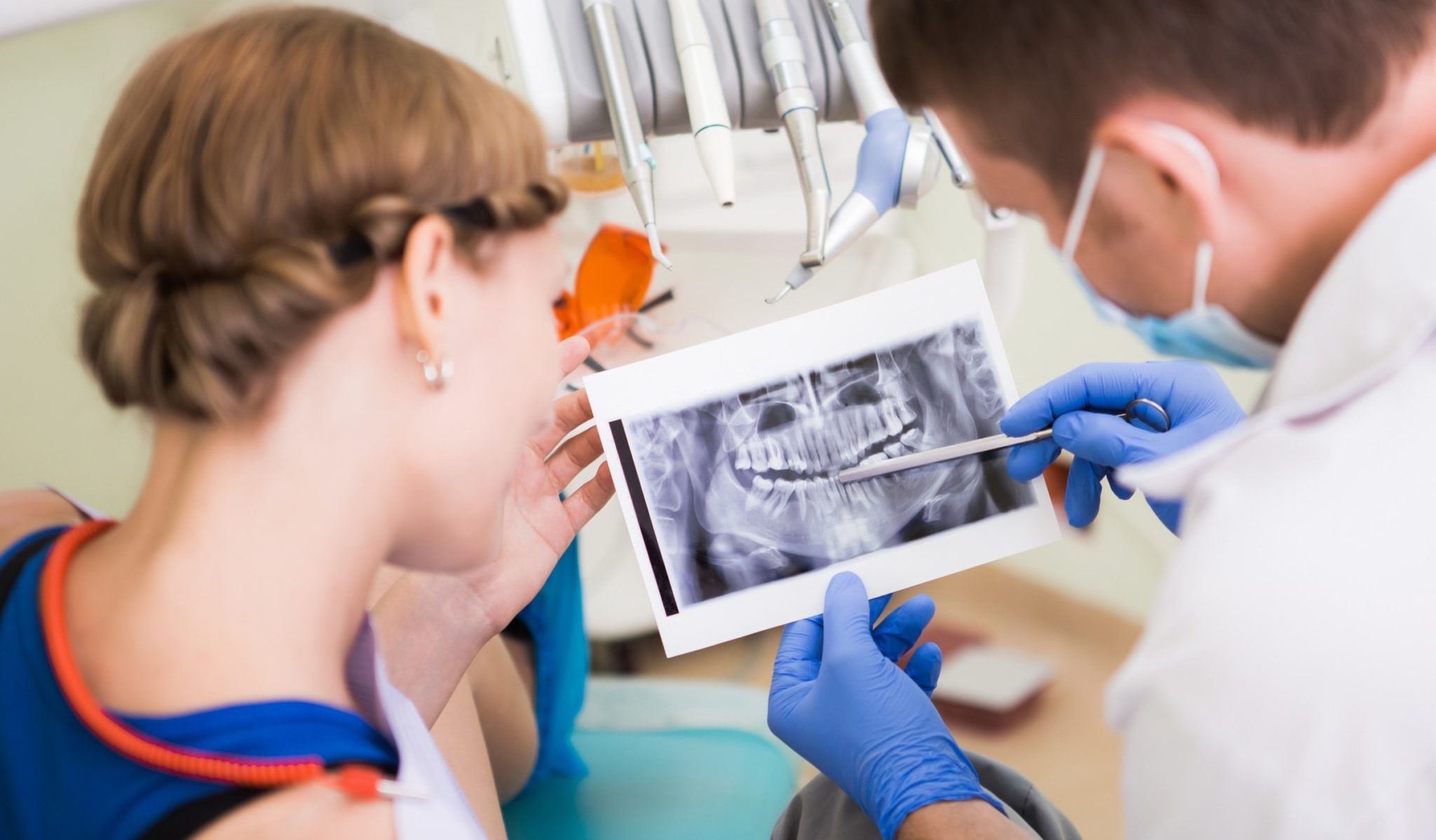
Before tooth extraction, treatment of dental operations, the doctor sends the patient to take a panoramic picture of the teeth or orthopantomogram (OPTG). This is an image of the bone and soft tissue of the dentition, according to it, the doctor can determine the condition of the dentition, periosteum, identify the true cause of the disease. The method is very useful, since an external examination gives only 50% information.
Content
What an orthopanorama photo shows
A picture of the OPTG of the teeth gives a clear picture of the state of the roots of the teeth and periosteum, which is convenient for a complete diagnosis and treatment.
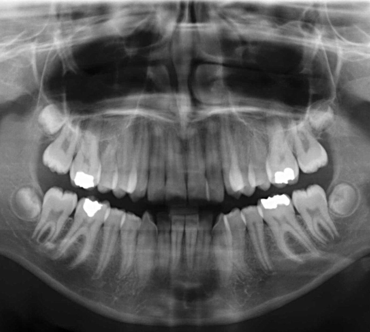
Caries in a panoramic shot
Using a panoramic photo you can identify:
- carious lesions hidden from the eyes;
- tumors, cysts, granulomas;
- pathology of the root system;
- lesions of the near-root space;
- condition of crowns and fillings;
- cracks, fractures, atrophic processes;
- horizontal and uncut teeth of wisdom;
- depth of gingival pockets;
- congenital pathology.
A panoramic picture of the jaw is an image produced by a digital camera. It can be printed as a photograph, viewed on a monitor and even sent to colleagues via the Internet.
When sent to OPTG
An orthopantomogram or panoramic picture of the teeth is done in the following cases:
- before root removal in order to prevent complications;
- in the treatment of periodontal disease to determine the group of the disease;
- in the diagnosis of jaw neoplasms;
- with flux;
- in orthopedic practice, for example, when you need to install braces;
- with traumatic damage to the jaw;
- with diagnoses of unknown etiology.
An x-ray helps the orthodontist to assess the condition of the bone and muscle systems of the oral cavity, to make timely treatment if any abnormalities are detected. And also to examine the maxillary sinuses with sinusitis and sinusitis.
Radiography is shown before tooth extraction, since the roots are visible on the film, the condition of the gums behind the dentition. Timely detection of pathology will help to avoid complications, so you can not pull out teeth without a preliminary panoramic picture, especially wisdom teeth.

Dental materials contain a composition that when shooting stands out in contrast. This is done so that you can track the shape of the filling material. A picture of the tooth on the film helps to choose the right treatment, allows you to make a complete picture of the condition of the mandibular canal, maxillary sinuses and nasal passages. The doctor has the opportunity to determine the position of the nasal septum. If it is curved, then the likelihood of chronic or allergic rhinosinusitis is high.
OCG gives a clear idea of the state of the dentition of the patient. Therefore, it is necessary for the qualitative diagnosis of diseases. With prolonged treatment, OPTG is done several times to monitor the healing process.
Procedure execution
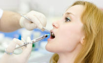
In vain, patients get scared when they hear that they are being sent to take a panoramic picture. This completely painless and quick procedure is much safer than x-rays. A digital orthopantomogram is performed within 30 seconds, after 5 minutes the image on the film will be ready.
There are no problems with orthopantomograms in large cities, as modern dental clinics are equipped with the necessary equipment. However, its high cost makes the examination unavailable in small towns, so many patients still cannot make an orthopantomogram for a full diagnosis of the disease.
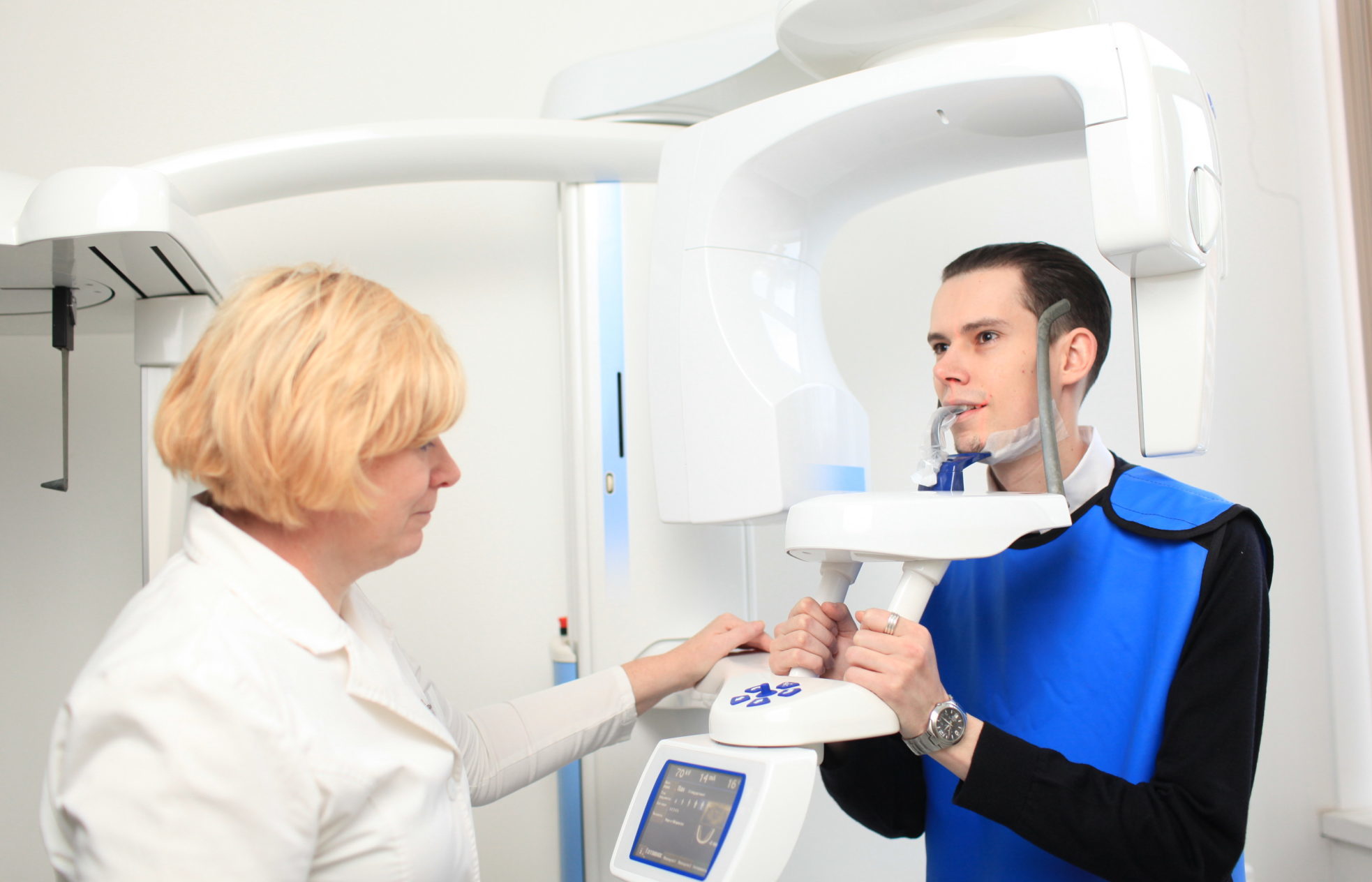
To obtain a tomogram, the patient needs:
- to free the head and neck from all metal objects and jewelry;
- located inside the orthopantomograph;
- put on an apron;
- hold a plastic tube in your mouth, closing your lips;
- grab the handles of the orthopantomograph;
- fix the pose for a while.
The device makes a circular revolution around the patient’s head and displays the finished image on the screen. In just a few minutes you can get a printed photo.
The main advantages of the procedure
Orthopantomography of teeth has many advantages:
- High security: the irradiating dose is much less than with radiography, which allows you to take a picture repeatedly. The procedure is harmless to children and the elderly.
- The speed of obtaining the final result.
- The ability to transfer images over the Internet.
- Image quality allows you to identify the disease in the early stages of development.
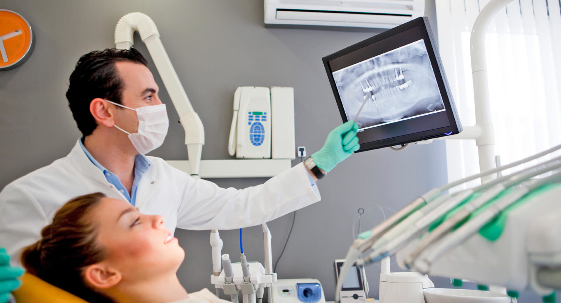
- Viewing on a monitor makes it easier for a doctor to diagnose and treat: an orthopanographic image can be enlarged for detailed examination of individual sites.
- The adjustable height of the emitter helps to adapt the device to any patient, whether it be a child or a disabled person in a stroller.
Individual flaws
Indications for orthopantomography have some limitations. Pregnant women are not recommended to undergo the procedure, like any other x-ray examination. Restrictions are also imposed on nursing mothers. In early infancy, only an orthodontist can send for an organized crime group.
The procedure is also contraindicated for people with severe diabetes mellitus, thyroid diseases, renal failure, allergy to iodine, and epilepsy.
Usually, the clinic doctor conducts a survey for any contraindication. If this does not happen, the patient should talk about deviations in health on his own.
How safe is OPTG?
The permissible dose of x-ray exposure for humans according to sanitary standards is 1.0 m3v. And the irradiation power with an orthopantomogram does not exceed 0.02 m3v; almost the same dose is received by an aircraft passenger during a flight.
This type of study is preferable to the X-ray method. All radiological procedures must be recorded in a medical record, records are kept so that the number of sessions does not exceed the permissible level.
Orthopantomogram interpretation
OPTG in dentistry is the ability to diagnose areas of the oral cavity hidden from the eyes: tooth roots, gum tissue. The doctor, looking at a survey picture of the teeth, will immediately determine the condition of the patient’s roots and jaw, detect a pathology and prescribe the correct treatment based on the result. A panoramic photo is invaluable for orthodontics, as it simplifies the diagnosis of many dental diseases.
For an ordinary patient, a panoramic picture is a complete mystery, but it can be decrypted. OPTG decryption is not so complicated. First you need to pay attention to the image on the film: the jaw should have a "laughing" look. If the dentition looks like a sad smile, this indicates a poor-quality image, looking at it, it is unlikely to be able to correctly diagnose the disease.
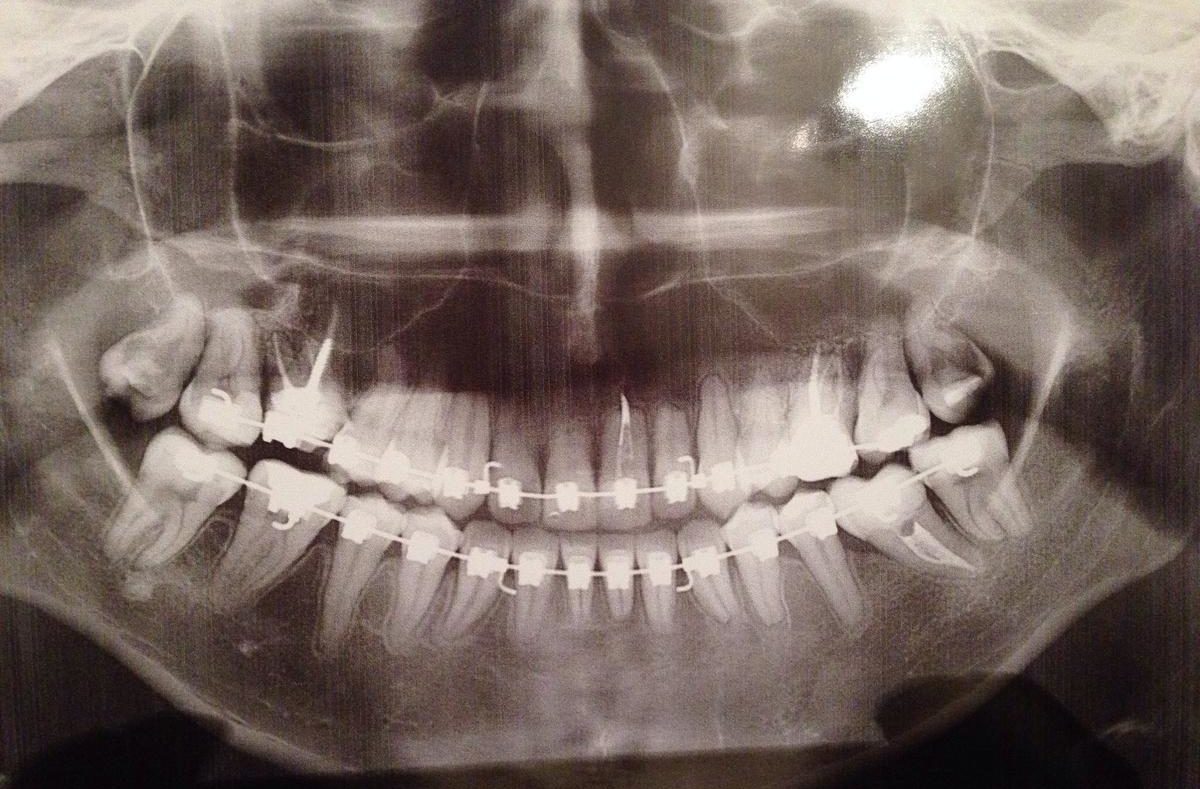
The orthopantomogram shows an image of the jaw, but not in mirror image. It must be considered as belonging to another person. This means that the image on the left side will be on the right, and the right on the left.
The classification of dentitions is deciphered in the form of numbering, but not the usual one. The fact is that the dentition in the picture is divided into four segments.The upper right is dozens, the upper left is twenty, thirty are the lower left, and the magpies are the lower right. Numbering starts with central incisors. The upper right central incisor is indicated by number 11. The wisdom tooth in the lower right row has the 48th number. Having learned this wisdom, you can pleasantly surprise the doctor by mentioning the numbering.
Children's orthopantomogram
An orthopantomogram or OPTG can be prescribed to children to get a picture of tooth growth. A periodic X-ray examination will help to identify abnormalities in the formation of dentitions, and take on time to correct the occlusion. Panoramic shots are allowed for children over 5 years old.
Postoperative OPTG
In modern dentistry clinics, a panoramic picture of the jaw is taken both before and after the operation. In the first case, it is necessary to obtain information about the condition of the jaws and to choose the optimal treatment method, and in the postoperative period to monitor the healing process.
Thanks to the overview picture, the dentist monitors changes in the patient’s health, notes the improvements, prevents complications in the healing process.
The cost of an X-ray examination is low, for example, in Moscow it will cost about 1 thousand rubles.