What teeth are called molars and premolars, anatomical features
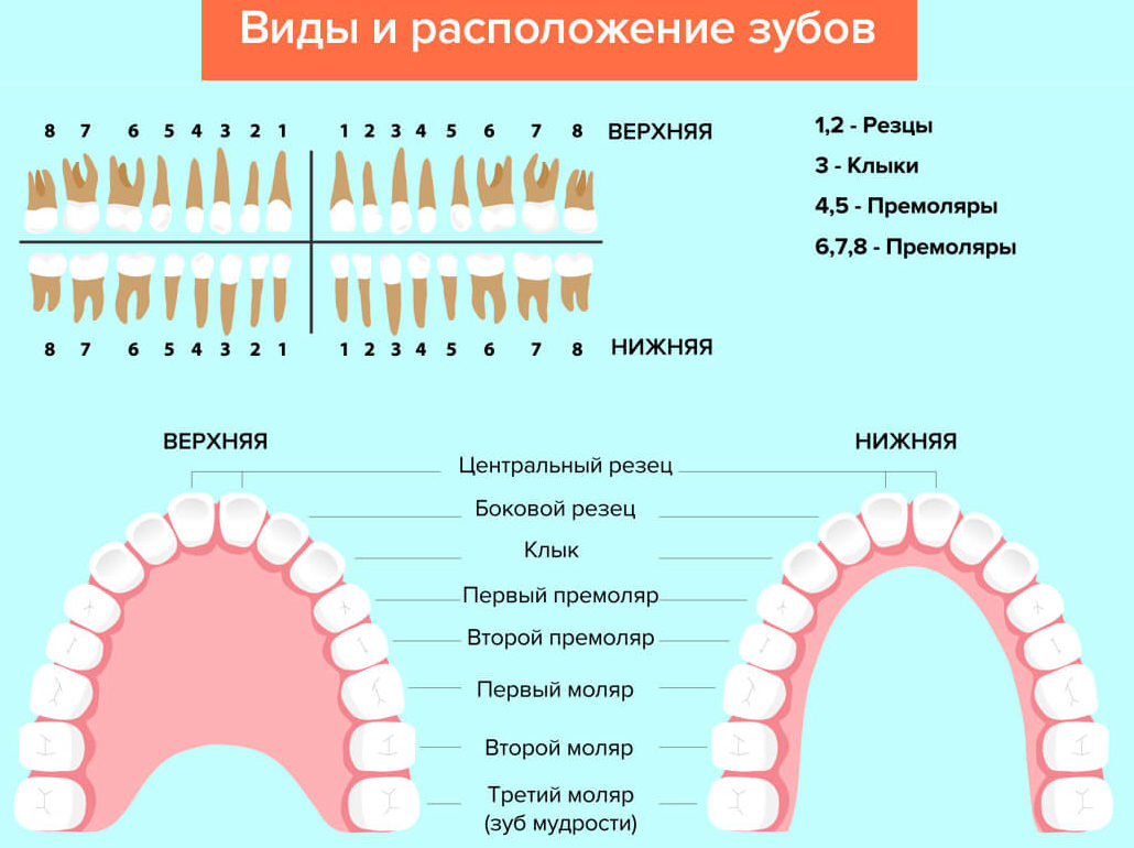
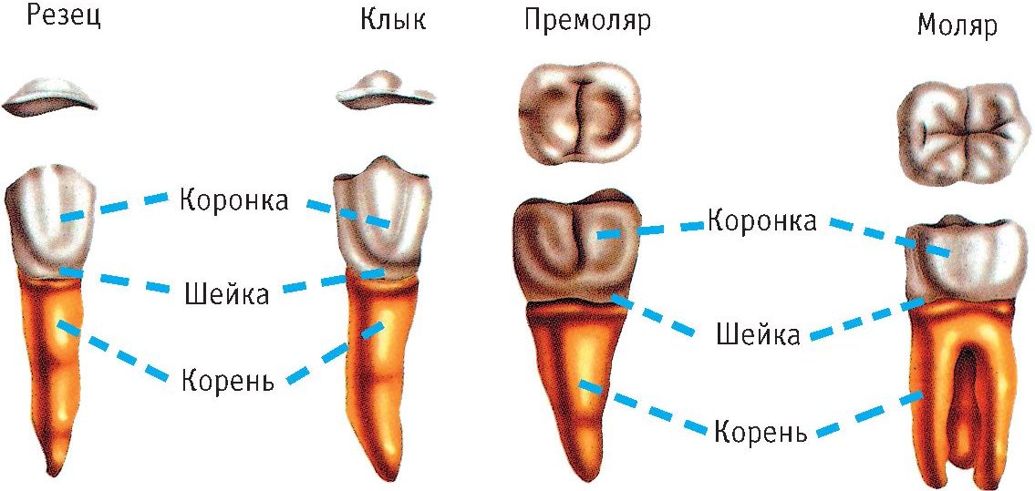
Fine, an adult should have 32 tooth units: sixteen on the lower and upper jaws. Their structure differs depending on the location and functional task. According to the same criteria, permanent teeth are divided into four types: molars intended for chewing and grinding food, fangs and incisors necessary for biting, tearing and holding, and premolars that perform all of these functions.
Content
Location and anatomical features of molars
Normally, every adult should have 12 molar units. They are located in pairs: three on the left and right sides of the upper and lower jaw. In adults, teeth from 6 to 8 are molar, in children - 4 and 5 teeth.
Molar teeth are the last elements in the jaw row. Their anatomical features are associated with functional purpose - grinding pieces of food.
The molars have the largest coronal part. This is due to the fact that when chewing they have the highest load - about 70 kg. Fangs experience a load not exceeding 40 kg.
Structural features of the lower and upper molars
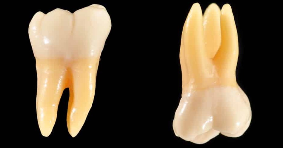
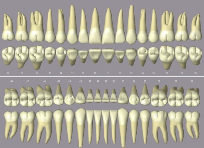
Lower molars usually have two roots and three channels. A characteristic feature of the upper is the presence of four channels and three roots. They are larger and have an anatomical structure different from the lower antagonists. A schematic photo of the teeth shows how different molars differ from each other.
The size of the crown of molar dental units varies from 7 to 9 mm. The chewing surface has a diamond shape with rounded corners. There are 4 tubercles on it, separated by three transverse grooves. There are usually three roots, in dentistry they are given the following names:
- palatine;
- buccal-mesial;
- buccal-distal.
The largest root is buccal-mesial, medium in size is palatine, and the shortest is buccal-distal. In rare cases, the upper molars can have 4 roots.
The lower large molars have a slightly smaller crown size. The number of tubercles on their chewing surface varies from 3 to 6. The medial and distal dental roots are parallel to each other. Often there is splicing of the roots.
Differences in the structure of molars under different serial numbers
Depending on the order of teething and location, the first, second and third molars are distinguished. Each subsequent molar tooth has smaller than the previous size of the crown and roots.
The first molars are the largest, they have the largest coronal surface area and the largest root size. The first large molar of the upper row has a more powerful root than its antagonist in the lower jaw. The crown of the first molar tooth in the lower jaw differs in a cubic shape and is slightly elongated along the jaw row.
The second molars on both jaws are smaller than the first. The upper second molars can have a crown of any shape, unlike the lower ones: they are characterized by a regular cubic shape and the presence of a clear cruciform groove dividing the crown surface into 4 tubercles.
Third molars are better known as wisdom teeth. They erupt at a conscious age and have no predecessors - milk molars.
Anatomical features of wisdom teeth:
- The size of the crown and the length of the root system can be different.
- Third molars located on top are smaller than those on the bottom. They can have one to five roots.
- On the crown there are usually three tubercles - two buccal and one lingual.
- The lower wisdom teeth are always larger than the upper. Usually they have two roots, but sometimes they grow together into one.
- The length of the roots is small, during growth they often deviate to the side.
What teeth are called premolars and features of their structure
Premolars are called 4 and 5 small molars located behind fangs. Dentists call them chewable. An adult has 8 small molars located in pairs on the right and left sides of both jaws.
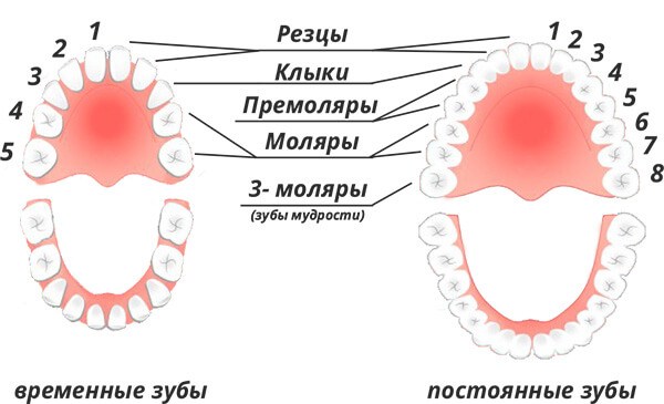
Premolars are not dairy, they erupt during the formation of a permanent bite. Children have milk molar teeth in their place, and premolar erupt after their loss (see photo). This is due to a lack of space on a small children's jaw.
Premolars belong to the transitional type of dental units - in terms of the size of the tooth crown and the structure of the root system, they are similar to fangs, but they resemble molars in terms of chewing surface area. The differences are clearly visible in the photo.
The main function of premolars is the same as that of canines - grabbing, tearing and crushing food. But due to the wider chewing surface, they are involved in the grinding of pieces of food.
The crowns of premolar teeth have a prismatic shape and two tubercles on the chewing surface. Upper premolar anatomically different from the lower:
- The upper ones are larger, have a more rounded barrel-shaped and two channels.
- Lower molars usually have one canal.
Features of the lower premolar
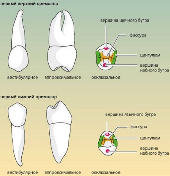
According to the anatomical features, the first premolar is similar to the neighboring canine. Its buccal surface is convex and longer than the palatine. The channel is usually one, but in rare cases there can be two.
The anatomical structure of the second premolar is similar to the second molar: the crown of the tooth is inclined inward, the size of the tubercles is approximately the same, between them is an enamel roller, separated from the faces by a horseshoe-shaped fissure. This structure allows him to withstand high chewing load and better grind food. The second premolar tooth unit has one cone-shaped, slightly flattened root.
Features of the upper premolar
The first premolar of the upper jaw, thanks to the pronounced vestibular tubercle, visually resembles a canine. The crown has a prismatic shape, the buccal tubercle is more pronounced than the palatine, between the tubercles there is a deep groove that does not reach the edges of the crown. Enamel rollers are located on the edge of the chewing surface. Root two - buccal and palatine.
The size of the palatine root exceeds the size of the buccal. Normally, they are divided in the apical region, but in dentistry there are cases of their separation in the middle and cervical region. There are usually two channels, in rare cases, one or three.
The second premolar is smaller than the previous one. Their structure is almost identical, except that the second has a less convex vestibular tubercle and one canal. A second maxillary premolar with two canals is a rare occurrence in less than a quarter of dental patients.
According to dental statistics, adult molars and premolars are particularly susceptible to caries. This is due to their inaccessibility during brushing and the complex structure of the tooth surface: the fissures covering it act as a favorable environment for the accumulation of pathogenic bacteria. Therefore, during procedures for oral hygiene, increased attention should be paid to cleaning the crown surface of teeth located at the end of the dentition.